Simple Squamous Epithelium Surface View Total Magnification
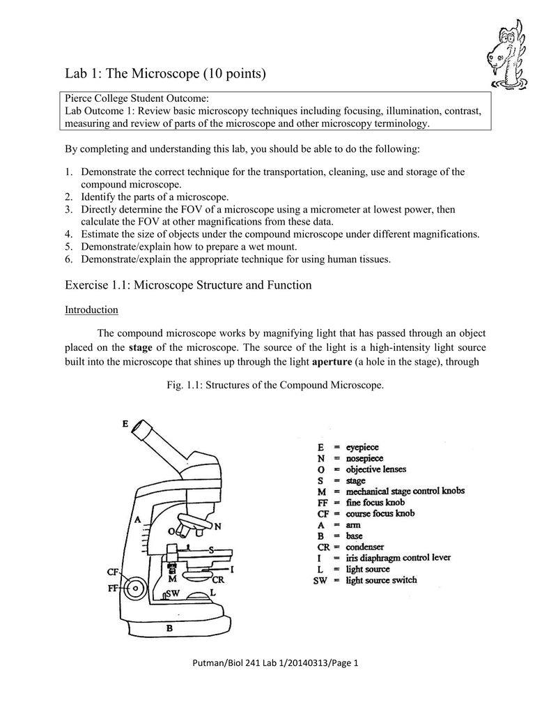
Objective Magnification 40X Total Magnification 400X TE=Transitional Epithelium N=Nucleus L=Lumen.

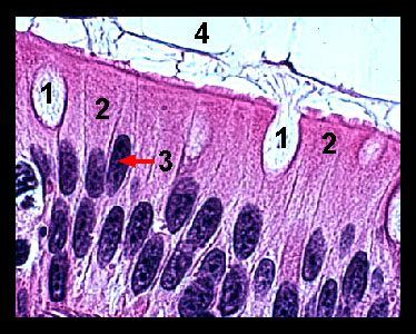
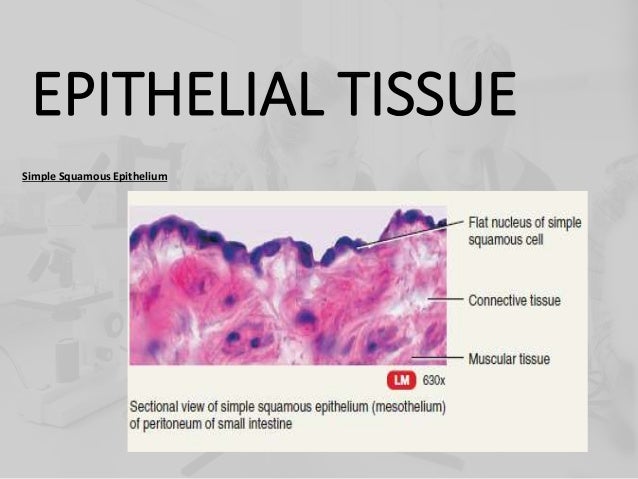
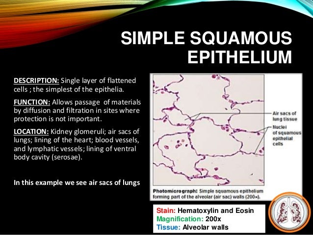
Simple squamous epithelium surface view total magnification. Muscularis mucosae (this is separate from the thicker layer of smooth muscle within the muscularis layer) Epithelium covers the inner surface of the digestive tract It starts as stratified squamous epithelium in the esophagus and changes to simple columnar epithelium in the stomach. Histology various type of epithelium 1 Simple Squamous Epithelium Composed of a single layer of flattened cells lying on a basement membrane In section only their flattened nuclei are often visible Function Provides a selective barrier which allows for filtration, passive diffusion and pinocytosis 2. The simple cuboidal epithelium is highlighted with a light blue outline This epithelium is easy to recognize because it is formed by cells that are equally tall and large, and by the presence of a roundish nucleus always positioned in the center of the cell Typically, the simple cuboidal epithelium lines ducts that contain fluids H&E X400.
Find the perfect Squamous Epithelium stock photos and editorial news pictures from Getty Images Select from premium Squamous Epithelium of the highest quality. Study their appearance and characteristics Correlate their physical structure with their function 6 simple squamous epithelium Drow a representation of Simple Squamous Epithelia at high magnification 7 Simple cuboidal epithelium Draw a representation of Simple cuboidal epithelium Label the apical surface, lumen and basement membrane 8. Same as 2, just a lower power magnification;.
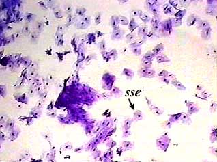
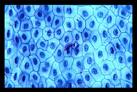
SSE Simple Squamous Epithelium L Lumen Wed, 02/07/18 2353 Permalink Please put the total Please put the total magnification in the description below your image, along with a key that defines what each label in (N= nucleus, etc) For the SSE label, try to bracket off a region of that type of epithelium, rather than just pointing. Simple squamous epithelium, (mesothelium) surface view, 250x shows shows squamous cells connected (sometimes called pavement epithelium), nuclei, cytoplasm, cell membrane this epithelium is found in the mesentery squamous epithelium stock pictures, royaltyfree photos & images. MESOTHELIUM (SIMPLE SQUAMOUS EPITHELIUM) view from surface Stained with silver nitrate 1 nucleus of cell 2 cell borders SIMPLE SQUAMOUS EPITHELIUM Stained with haematoxylin and eosin Nuclei of the epithelial cells are shown with an arrow SIMPLE CUBOIDAL EPITHELIUM.
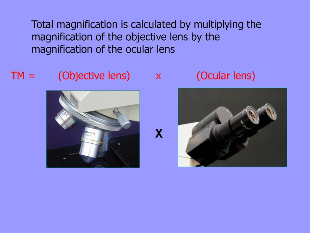
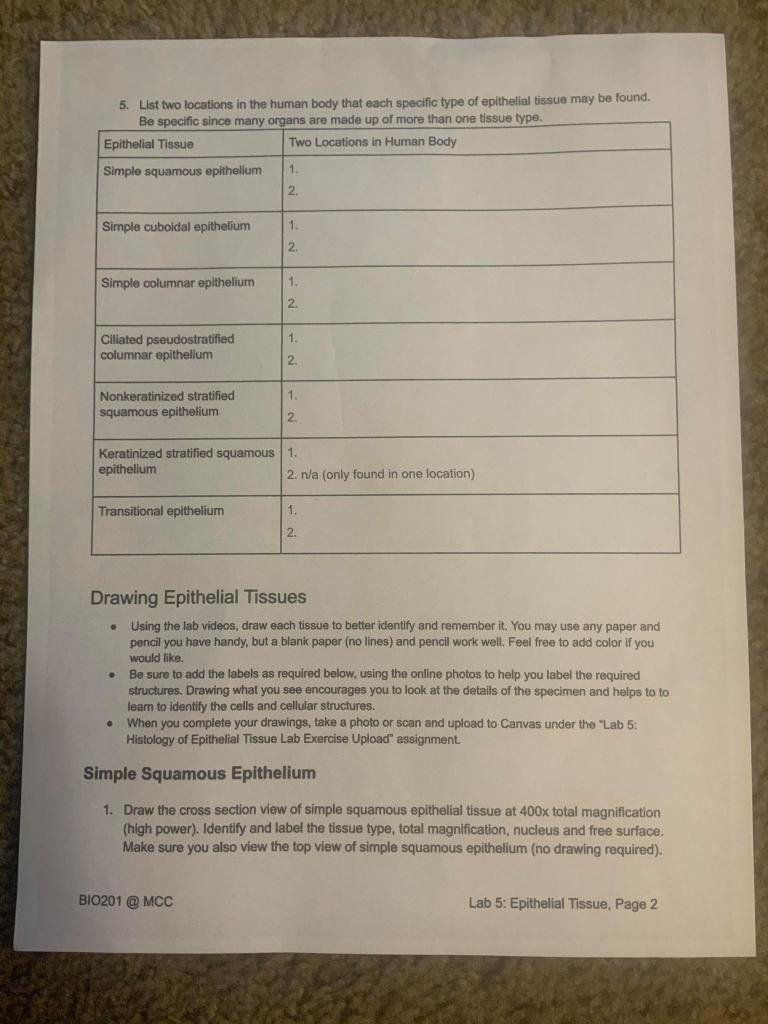
•Total magnification calculated by multiplying the two •Can view surface features only •Maximum resolution of about 10 nm (001 μm) Simple squamous epithelium Most delicate epithelium (one layer thick) Functions include absorption, diffusion, reduction of. Find the perfect Squamous Epithelium stock photos and editorial news pictures from Getty Images Select from premium Squamous Epithelium of the highest quality. Cells enlarge as they approach surface of lumen *typical "pear" shape of this epithelium.
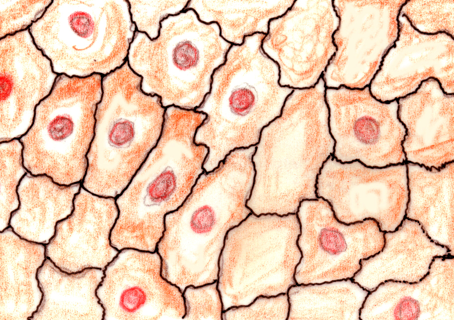
Squamous cells forming the wall of an alveolus;. Simple squamous epithelium is the tissue that creates from one layer of squamous cells which line surfaces The squamous cells are thin, large, and flat, and consisting of around nucleus These tissues have polarity like other epithelial cells and consist of a distinct apical surface with special membrane proteins. Simple Squamous Epithelium Definition Simple squamous epithelia are tissues formed from one layer of squamous cells that line surfaces Squamous cells are large, thin, and flat and contain a rounded nucleus Like other epithelial cells, they have polarity and contain a distinct apical surface with specialized membrane proteins.
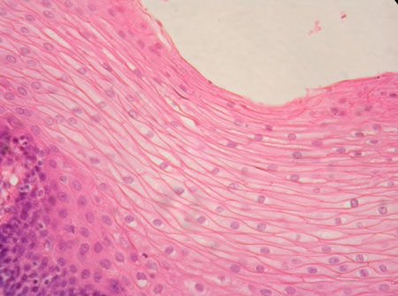
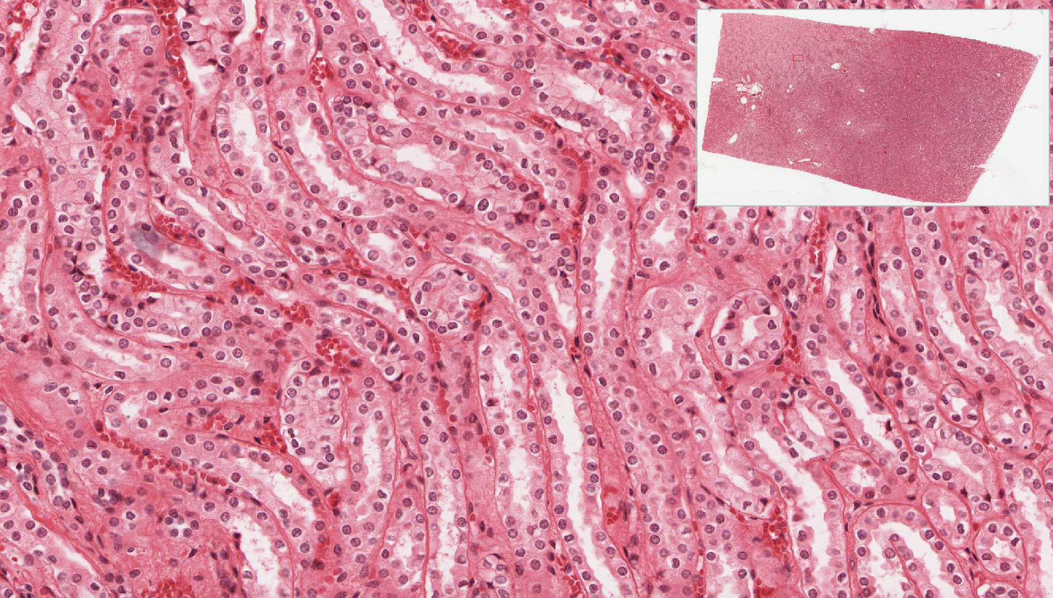
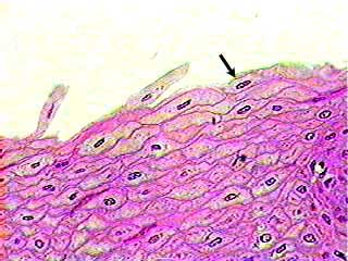
A stratified squamous epithelium is made up of a number of layers and the cells of the outer layers are flat (squamous) In the deepest layer new cells are produced by the division of stem cells Cells are gradually pushed toward the surface by the production of newer cells. The simple squamous epithelial lining of the blood vessel is a single layer of flat, or squamous, cells The basement membrane is so thin it is undectable at this magnification The nucleus is usually the only cellular structure visible at this magnification. Simple squamous epithelial cells of the kidney Bowman’s capsule, low magnification Bowman's capsule of a kidney corpuscle The parietal layer (the outer edge of the capsule) is a simple squamous epithellium The capillaries inside the capsule (the glomerulus) are also lined by a squamous epithelium, the endothelium It's hard to see.
11 Practice MidTerm Exam Key to Practice Benchtop Practical Pictures of some models used on the 11 midterm exam Things you should know about the exam Lab 1 parts of a m. Epithelium Web Lab A Squamous Epithelium 1 Isolated Squamous Epithelial Cells in face view (Cheek Cells) Draw one Squamous epithelium cell on high power and label the nucleus, cytoplasm and cell membrane, Be sure to label the magnification * low power * medium power * high power 2 Simple Squamous Epithelium (cross section/transverse) These slides will show a cross section of the small. A Simple columnar epithelium Slide 29 (small intestine) View Virtual Slide Slide 176 40x (colon, H&E) View Virtual Slide Remember that epithelia line or cover surfaces In slide 29 and slide 176, this type of epithelium lines the luminal (mucosal) surface of the small and large intestines, respectively Refer to the diagram at the end of this chapter for the tissue orientation and consult.
A stratified epithelium is more than one layer of cells thick A pseudostratified epithelium is really a specialized form of a simple epithelium in which there appears at first glance to be more than one layer of epithelial cells, but a closer inspection reveals that each cell in the layer actually extends to the basolateral surface of the. Squamous cells forming the wall of an alveolus;. 11 Practice MidTerm Exam Key to Practice Benchtop Practical Pictures of some models used on the 11 midterm exam Things you should know about the exam Lab 1 parts of a m.
Simple Squamous Epithelium Definition Simple squamous epithelia are tissues formed from one layer of squamous cells that line surfaces Squamous cells are large, thin, and flat and contain a rounded nucleus Like other epithelial cells, they have polarity and contain a distinct apical surface with specialized membrane proteins. 21 Simple squamous epithelium (Top view) 22 Simple cuboidal epithelium (Collecting tubules) Example 10x ocular X 10x objective=100x total magnification Note A slide will be given to you fix the slide on the stage and focus and observe Covering “surface” epithelium Glandular epithelium Neuro_epithium. A Simple columnar epithelium Slide 29 (small intestine) View Virtual Slide Slide 176 40x (colon, H&E) View Virtual Slide Remember that epithelia line or cover surfaces In slide 29 and slide 176, this type of epithelium lines the luminal (mucosal) surface of the small and large intestines, respectively Refer to the diagram at the end of this chapter for the tissue orientation and consult.
Characteristics Epithelial tissue is classified based on the shape of the cells at the apical surface and the number of layers of cells This tissue consists of 1 layer of cells that have a scalelike, flattened, or squished shape. An epithelium is the type of tissue that covers surfaces, usually the linings of hollow organs in the body, or in the case of the skin, the outer surface of the body In many cases, adjacent epithelial cells are linked by tight junctions so that the epithelium forms a barrier that regulates the movement or substances across it (see the web page on Epithelial Transport). The simple cuboidal epithelium is highlighted with a light blue outline This epithelium is easy to recognize because it is formed by cells that are equally tall and large, and by the presence of a roundish nucleus always positioned in the center of the cell Typically, the simple cuboidal epithelium lines ducts that contain fluids H&E X400.
Microscopic view of slide of simple cuboidal epithelium in 4x, 10x & 40x magnification. Figure 41 ¦ Simple squamous epithelium surface view of peritoneal mesothelium figure 42 ¦ Simple squamous epithelium peritoneal mesothelium surrounding small intestine (transverse section) figure 43 ¦ Different epithelial types in the kidney cortex figure 44 ¦ Simple columnar epithelium surface of stomach. Most epithelial cells are separated from the connective tissue by a sheet of extracellular material called the basal lamina This structure is visible only with the electron microscope, where it appears as a dense layer, –100 nm thick, consisting of a delicate network of very fine fibrils (lamina densa) In addition, basal laminae may have an electronlucent layer on one or both sides of.
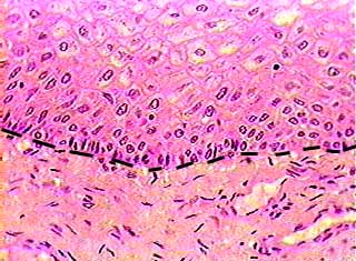
Characteristics Epithelial tissue is classified based on the shape of the cells at the apical surface and the number of layers of cells This tissue consists of 1 layer of cells that have a scalelike, flattened, or squished shape. A simple squamous epithelium is a single layer of flat cells in contact with the basal lamina (one of the two layers of the basement membrane) of the epithelium This type of epithelium is often permeable and occurs where small molecules need to pass quickly through membranes via filtration or diffusionSimple squamous epithelia are found in capillaries, alveoli, glomeruli, and other tissues. This is nonkeratinized, stratified squamous epithelium The epithelium fills most of the screen the tissue stretches from the green apical surface to the blue basement membrane Note the basement side are both cuboidal in shape and very healthy looking Alternatively, the apical cells, have flattened, squamous quality.
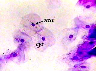
Simple Squamous Epithelium Definition Simple squamous epithelia are tissues formed from one layer of squamous cells that line surfaces Squamous cells are large, thin, and flat and contain a rounded nucleus Like other epithelial cells, they have polarity and contain a distinct apical surface with specialized membrane proteins. High magnification of transitional epithelium of urinary bladder *note multicellular nature of epithelium;. Simple squamous epithelium, isolated (400x) Buccal mucosal In the center of this image are two simple squamous epithelial cells that are still attached to each other Notice that the location of the nucleus (nuc) is in the center of the cell It is surrounded by the much paler cytoplasm (cyt).
Total magnification answer passageway that the apical surface of an epithelial tube is exposed to the inside of question basolateral surfaces the simple squamous epithelium lining the inner surface of the heart and all blood vessels question stratified squamous epithelium. Simple epithelium has only one cell layer where every cell is in direct contact with the underlying basement membrane Generally, this type of epithelium is found inside the body probably due to the fragile nature and forms the lining of the body cavities, blood and lymph vessels, heart and respiratory system Being a thin layer has the physiological advantage of faster absorption and. An epithelium is the type of tissue that covers surfaces, usually the linings of hollow organs in the body, or in the case of the skin, the outer surface of the body In many cases, adjacent epithelial cells are linked by tight junctions so that the epithelium forms a barrier that regulates the movement or substances across it (see the web page on Epithelial Transport).
Because simple squamous tissue is so thin, it's not a great layer of protection Its tissuethin surface could tear easily and doesn't shield the tissue underneath However, the squamous cells' thin structure means simple squamous epithelial tissue is great for helping to absorb, diffuse and release substances. SIMPLE SQUAMOUS EPITHELIUM (Mesothelium) Surface View, 250X Shows shows squamous cells connected (sometimes called pavement epithelium), nuclei, cytoplasm, cell membrane This epithelium is found in the mesentery stock photo. Simple epithelium has only one cell layer where every cell is in direct contact with the underlying basement membrane Generally, this type of epithelium is found inside the body probably due to the fragile nature and forms the lining of the body cavities, blood and lymph vessels, heart and respiratory system Being a thin layer has the physiological advantage of faster absorption and.
Simple squamous epithelium forms the inside walls of blood vessels (endothelium), the wall of Bowman's capsule of the kidney, the lining of the body cavity and viscera (parietal and visceral peritoneum) and the walls of the air sacs (alveoli) and respiratory ducts of the lung Surface view Lab2 23 19 Simple cuboidal epithelium model. Epithelium (/ ˌ ɛ p ɪ ˈ θ iː l i ə m /) is one of the four basic types of animal tissue, along with connective tissue, muscle tissue and nervous tissueIt is a thin, continuous, protective layer of cellsEpithelial tissues line the outer surfaces of organs and blood vessels throughout the body, as well as the inner surfaces of cavities in many internal organs. Locations of simple squamous epithelium alveoli of lungs, walls of capillaries, linings of blood vessels and ventral body cavity functions of simple cuboidal epithelium.
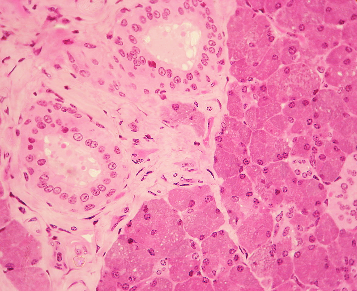
(Total Magnification) Specific Cells are not labeled within each tissue, remember that many of Simple Columnar Epithelium 100x Simple Columnar Epithelium 400x Simple Cuboidal 40x Simple Squamous (bowman’s Capsules) 400x Stratified Squamous 40x Stratified Squamous 100x. Simple squamous epithelium, cs (400X) thin section Kidney cortex The arrows in the image point to the nuclei of simple squamous epithelial cells This image was made from a thin section of the kidney at the same magnification as the previous image (400X) It is about onefifth to onetenth the thickness of the slides used to make the top. Low Magnification Survey the renal cortex with 10x objective and find a satisfactory renal corpuscle Center the corpuscle in your field of view The simple squamous epithelium is too thin to resolve, so rotate the high magnification objective into position and continue your study.
A simple squamous epithelium is a single layer of flat cells in contact with the basal lamina (one of the two layers of the basement membrane) of the epithelium This type of epithelium is often permeable and occurs where small molecules need to pass quickly through membranes via filtration or diffusionSimple squamous epithelia are found in capillaries, alveoli, glomeruli, and other tissues. Simple squamous epithelial cells of the kidney Bowman’s capsule, low magnification Bowman's capsule of a kidney corpuscle The parietal layer (the outer edge of the capsule) is a simple squamous epithellium The capillaries inside the capsule (the glomerulus) are also lined by a squamous epithelium, the endothelium It's hard to see. SIMPLE SQUAMOUS EPITHELIUM (Mesothelium) Surface View, 250X Shows shows squamous cells connected (sometimes called pavement epithelium), nuclei, cytoplasm, cell membrane This epithelium is found in the mesentery stock photo.
SIMPLE SQUAMOUS EPITHELIUM (Mesothelium) Surface View, 250X Shows shows squamous cells connected (sometimes called pavement epithelium), nuclei, cytoplasm, cell membrane This epithelium is found in the mesentery stock photo. LS, lumenal surface Survey the slide with the scanning objective Locate the lumenal surface of the blood vessel The lumenal surface of the vessel is lined by a simple squamous epithelium called endothelium The endothelium is so thin, it is virtually unresolvable at this magnification. Simple squamous epithelium Simple squamous epithelia consist of a single layer of flattened cells This type of epithelia lines the inner surface of all blood vessels (endothelium), forms the wall of alveolar sacs in the lung and lines the body cavities (mesothelium).
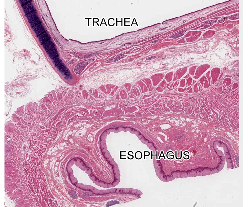
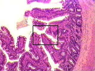
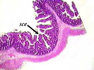
The surface of the intestine exposed to the peritoneal cavity (PC) is covered with a simple squamous epithelium known as a mesothelium The next slide is taken from the boxed area This high power view of the abluminal (the side away from the lumen) wall of the gut illustrates the single layer of squamous cells (a simple, squamous epithelium. Because simple squamous tissue is so thin, it's not a great layer of protection Its tissuethin surface could tear easily and doesn't shield the tissue underneath However, the squamous cells' thin structure means simple squamous epithelial tissue is great for helping to absorb, diffuse and release substances. A stratified epithelium is more than one layer of cells thick A pseudostratified epithelium is really a specialized form of a simple epithelium in which there appears at first glance to be more than one layer of epithelial cells, but a closer inspection reveals that each cell in the layer actually extends to the basolateral surface of the.
A Simple columnar epithelium Slide 29 (small intestine) View Virtual Slide Slide 176 40x (colon, H&E) View Virtual Slide Remember that epithelia line or cover surfaces In slide 29 and slide 176, this type of epithelium lines the luminal (mucosal) surface of the small and large intestines, respectively Refer to the diagram at the end of this chapter for the tissue orientation and consult. Same as 2, just a lower power magnification;. (Total Magnification) Specific Cells are not labeled within each tissue, remember that many of Simple Columnar Epithelium 100x Simple Columnar Epithelium 400x Simple Cuboidal 40x Simple Squamous (bowman’s Capsules) 400x Stratified Squamous 40x Stratified Squamous 100x.
Q Tbn And9gcq 2liziqu0pgql01yxvsbmyflq Jcb7aq0hkvnirqmwuy9t4j Usqp Cau
Www Drcroes Com Uploads 8 1 2 8 Anatomy Tissue Review Slides Part I Pdf
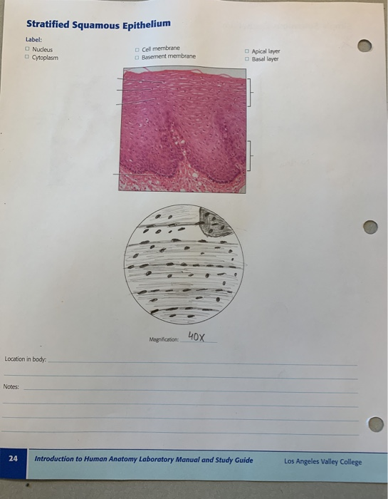
Stratified Squamous Epithelium
Simple Squamous Epithelium Surface View Total Magnification のギャラリー
Http Shammer Irsc Weebly Com Uploads 1 9 8 6 Histology Pdf

Lab Reporrt 5 Docx Lab Report 5 Name Angel Liana Marie Cervantes Epithelial Tissue Date Trial Number 1 Identifying Code Frog Skin Tissue Type Simple Course Hero
Solved Simple Squamous Epithelium Label Nucleus Cytoplas Chegg Com
Page 2 Epithelial Cells High Resolution Stock Photography And Images Alamy

Photomicrograph Of The Excised Uvula Mass Showing Normal Stratified Download Scientific Diagram

Histolab2 Htm
Exercise 4 Epithelium
Lab 2 Epithelial Tissue Histology
Epithelial Tissue Histology

Lab 2 Epithelial Tissue Histology

Lab 2 Epithelial Tissue Histology

Lab 2 Epithelial Tissue Histology
Simple Squamous Epithelium Isolated

Lab 2 Epithelial Tissue Histology
Histology Tutor
Http Histologyguide Org About Us Sorenson Atlas Of Human Histology Chapters 1 And 14 Pdf

Lab Manual Exercise 1
Http Www Tycmhoffman Com Commonfiles Bio354 Laboratory02 Pdf
Simple Squamous Epithelium Isolated
Q Tbn And9gctzu7fd7f 63uvsuhii Iwznc54mlzqmr8fup1orkbsues3tngw Usqp Cau
Stratified Cuboidal Epithelium Definition And Function Biology Dictionary

Lab Manual Exercise 1

Examining Epithelial Tissue Under The Microscope Human Anatomy And Physiology Lab Bsb 141
Epithelial Tissue Histology

Histolab2 Htm
Www Drcroes Com Uploads 8 1 2 8 Anatomy Tissue Review Slides Part I Pdf
Week 2 Microscopic Anatomy Ppt Download
Simple Squamous Epithelium Isolated
Solved Biol 123 Molecular And Cellular Biology Lab Score Chegg Com
Examining Epithelial Tissue Under The Microscope Human Anatomy And Physiology Lab Bsb 141
Solved Simple Squamous Epithelium Label Nucleus Cytoplas Chegg Com
Solved Simple Squamous Epithelium Label Nucleus Cytoplas Chegg Com

Stratified Squamous Non Keratinized Epithelium
Bisc 101 Textbook Notes Fall 12 Cardiac Muscle Blood Plasma Optical Microscope

Using The Rule Of Nines Answer Each Of The Questions That Follow R T Baraka Was Course Hero

Biol 160 Lab Practical 1 Flashcards For General Biology Flashcards Quizlet

Lab Manual Exercise 1
1 5 Microscopy Biology Libretexts
Stratified Squamous Non Keratinized Epithelium
Lab 2 Microscopy And The Study Of Tissues Zoo Lab Uw La Crosse

Chapter 02 Page 21 Histologyolm 4 0
Http Downloads Lww Com Wolterskluwer Vitalstream Com Sample Content Mcconnell Samples Chapter 07 Lab Manual Pdf

Stratified Squamous Epithelium

Hematoxylin And Eosin Histology Of Human Tissue Engineered Colon A Download Scientific Diagram

Histology Docx Epithelium Tissue Biology
Epithelial Tissue Histology
Columnar Epithelial High Resolution Stock Photography And Images Alamy

Lab Reporrt 5 Docx Lab Report 5 Name Angel Liana Marie Cervantes Epithelial Tissue Date Trial Number 1 Identifying Code Frog Skin Tissue Type Simple Course Hero

Assignment For The Science Biology Docsity

Chapter 1 Page 9 Histologyolm
Histologylab Ctl Columbia Edu Histologylabmanual Pdf
Www Palmbeachstate Edu Slc Documents ndpch04lecturepearson Pdf
Www Palmbeachstate Edu Slc Documents ndpch04lecturepearson Pdf
Http Fac Ksu Edu Sa Sites Default Files Mlzm Lmly 103 Khyn Pdf
Www Augusta Edu Scimath Biology Docs Animal Tissues F17 Pdf

Examining Epithelial Tissue Under The Microscope Human Anatomy And Physiology Lab Bsb 141

Histology Study Of Tissues Ppt Download
Bj9k0f L Ookam
2 3 Instruments Of Microscopy Biology Libretexts

Examining Epithelial Tissue Under The Microscope Human Anatomy And Physiology Lab Bsb 141

Biol 160 Lab Practical 1 Flashcards For General Biology Flashcards Quizlet
Instruments Of Microscopy Microbiology
2 3 Instruments Of Microscopy Microbiology Canadian Edition
Solved Tissues Lab Guidelines Draw Clearly And Carefully Chegg Com
Lab 1 The Microscope 10 Points Manualzz
I E All
Epithelial Tissue Histology

Simple Squmous Epithelium C S

Chapter 02 Page 21 Histologyolm 4 0

A P 242 Lab 2 Microscope Cells And Epithelial Tissues

Chapter 1 Review And Flashcards Quizlet

Examining Epithelial Tissue Under The Microscope Human Anatomy And Physiology Lab Bsb 141
Simple Columnar Epithelium

Chapter 1 Page 6 Histologyolm
Stratified Cuboidal Epithelium Wikipedia

Simple Squamous Epithelium Nuclei Cytoplasm Cell Membrane This Epithelium Is Found In The Mesentery High Res Stock Photo Getty Images
Histologylab Ctl Columbia Edu Histologylabmanual Pdf

Lab Manual Exercise 1
Solved Tissues Lab Guidelines Draw Clearly And Carefully Chegg Com
Q Tbn And9gct7egvdok63lxw Suvrh1qvsnsvgadjy3bxyyazzgreqvz96p Usqp Cau

An Anti Il 13 Antibody Reverses Epithelial Mesenchymal Transition Biomarkers In Eosinophilic Esophagitis Phase 2 Trial Results Journal Of Allergy And Clinical Immunology

Histolab2 Htm
Q Tbn And9gcskx04lcbk Fptlwmgrku8pxxpte1lwkn873papzge Ysfruacx Usqp Cau

Lab 3 Use Of The Microscope Pdf Free Download
Lab 2 Microscopy And The Study Of Tissues Zoo Lab Uw La Crosse
Histolab2 Htm

Teratomatous Components Of Keratinizing Stratified Squamous Epithelium Download Scientific Diagram
I E All
Microscopy And Cell Diversity

Lab 1 The Microscope 10 Points Pdf Free Download
Lab 2 Microscopy And The Study Of Tissues Zoo Lab Uw La Crosse
Stratified Squamous Non Keratinized Epithelium

Simple Squamous Epithelium 40x Annotated Histology
Solved Clinical Application Patches Of Material Containin Chegg Com
Simple Columnar Epithelium
Microscopes
Academicworks Cuny Edu Cgi Viewcontent Cgi Article 1015 Context Qc Oers
Biology 111 Introduction To The Microscope And
Frontiers Asymptomatic Bacterial Vaginosis Is Associated With Depletion Of Mature Superficial Cells Shed From The Vaginal Epithelium Cellular And Infection Microbiology
Www Drcroes Com Uploads 8 1 2 8 Anatomy Tissue Review Slides Part I Pdf