Simple Squamous Epithelium Surface View 1000x
Simple squamous epithelium is the tissue that creates from one layer of squamous cells which line surfaces The squamous cells are thin, large, and flat, and consisting of around nucleus These tissues have polarity like other epithelial cells and consist of a distinct apical surface with special membrane proteins.
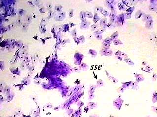
Simple squamous epithelium surface view 1000x. Lining body cavities and free surfaces of organs found in those cavities Kidney Name the tissue type?. MESOTHELIUMSIMPLE SQUAMOUS EPITHELIUM, Surface View, 400X, also called "pavement epithelium" These are very flattened cells showing a nucleus, cytoplasm and cell membrane This epithelium forms a thin and often permeable membrane Mesothelium is a special type of simple squamous epithelium that is found in serous membranes lining the pleural. The cheek individual squamous cells from the surface of the epithelium can be removed These surface cells are squamous (flat) in shape These cells are part of the nonkeratinized stratified squamous epithelium, but are large and are primarily going to be positioned such that they will be viewed from the “topside” rather than a side view.
Simple squamous (skwa’mus) epithelium consists of thin, flat cells that have an irregular outline and a flat, centrally located nucleus In a surface view, the cells somewhat resemble tiles arranged in a mosaic pattern. The main classifications of epithelium are simple and stratified, each one being further divided into several subtypes according to two main factors cell shape and apical surface specialization This article will describe stratified (multilayered) epithelium, focusing on its general characteristics and each major subtype. Simple Epithelium 1 Simple squamous epithelium is composed of one layer of squamous cells that rests on a basement membrane A surface view shows that the cells have a polygonal outline A side view shows that cells are extremely flattened with flattened nuclei Sites It is characteristically thin to allow material transport across it.
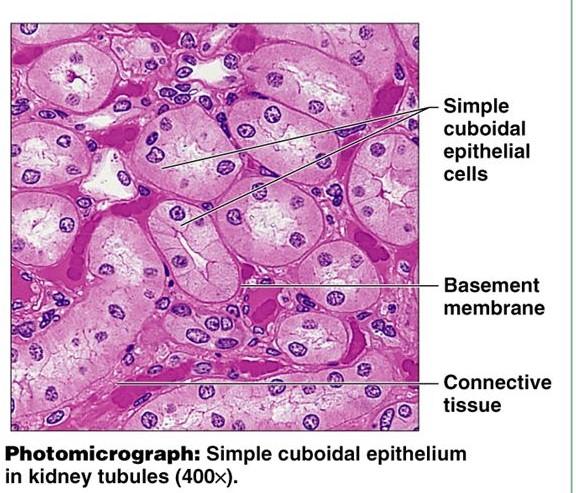
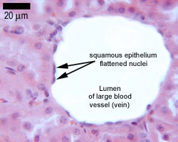
Simple squamous epithelium kidney long section 1000x Simple squamous epithelium venule longitudinal section 1000x Simple squamous epithelium mesothelium spread preparation 400x Simple cuboidal epithelium kidney longitudinal section 100x. Because simple squamous tissue is so thin, it's not a great layer of protection Its tissuethin surface could tear easily and doesn't shield the tissue underneath However, the squamous cells' thin structure means simple squamous epithelial tissue is great for helping to absorb, diffuse and release substances. The mesothelium is a simple squamous epithelium that forms the surface layer of the serous membrane that lines body cavities and internal organs Its primary function is to provide a smooth and protective surface Mesothelial cells are squamous epithelial cells that secrete a fluid that lubricates the mesothelium.
Simple squamous (skwa’mus) epithelium consists of thin, flat cells that have an irregular outline and a flat, centrally located nucleus In a surface view, the cells somewhat resemble tiles arranged in a mosaic pattern. View Available Hint(s) Simple squamous cells exist only as individual, independent cells and not as part of a population of simple squarous cells Simple squamous epithelium is always found wedged between other tissue types Simple squamous epithelium is found next to free space called the lumen Simple squamous epithelium consists of multiple. Chief examples are (1) endothelium which covers the internal surface of heart, blood vessels and lymph vessels, (2) mesothelium which lines the pericardial, pleural and peritoneal cavities, and (3) the lining epithelium of the alveoli of the lung.
Simple squamous epithelium ;. They may consist of a single layer of cells (simple) or more than one layer (stratified);. Simple Squamous Epithelium, 40X Submitted by ipolunina on Wed, 02/21/18 1338 Read more about Simple Squamous Epithelium, 40X;.
The mesothelium is a simple squamous epithelium that forms the surface layer of the serous membrane that lines body cavities and internal organs Its primary function is to provide a smooth and protective surface Mesothelial cells are squamous epithelial cells that secrete a fluid that lubricates the mesothelium. Simple squamous epithelium is of common occurrence in the body;. Simple squamous epithelium – a single layer of thin flattened cells This type of epithelium forms thin delicate sheets of cells through which molecules can easily pass (diffusion, filtration) Contiguous squamous epithelial cells also provide a smooth flat surface over which fluids and other tissues can move with low friction.
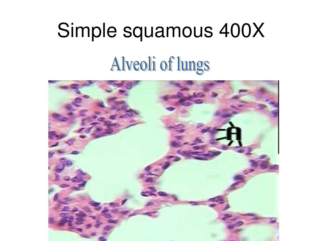
The mesothelium is a simple squamous epithelium that forms the surface layer of the serous membrane that lines body cavities and internal organs Its primary function is to provide a smooth and protective surface Mesothelial cells are squamous epithelial cells that secrete a fluid that lubricates the mesothelium. Simple Squamous Epithelium (1000x) Simple Squamous Epithelium (400x) Simple Squamous Epithelium (Spread 400x) Simple Squamous Epithelium (Venule) Stratified Cuboidal Epithelium Nonkeratinized Stratified Squamous Epithelium (100x) Keratinized Stratified Squamous Epithelium (Thick Skin 400x). The thinness of the barrier separating incoming air from capillary blood facilitates gaseous exchange 1000x The walls of adjacent alveoli abut to form interalveolar septa Each septum is lined by a simple squamous epithelium;.
The mesothelium is a simple squamous epithelium that forms the surface layer of the serous membrane that lines body cavities and internal organs Its primary function is to provide a smooth and protective surface Mesothelial cells are squamous epithelial cells that secrete a fluid that lubricates the mesothelium. Chief examples are (1) endothelium which covers the internal surface of heart, blood vessels and lymph vessels, (2) mesothelium which lines the pericardial, pleural and peritoneal cavities, and (3) the lining epithelium of the alveoli of the lung. Caitlin Kenney Date January 25, 21 In the alveoli, simple squamous epithelium helps diffuse gases in and out of the blood stream Simple squamous epithelium is a type of epithelial tissue characterized by a single layer of squamous epithelial cellsEpithelium lines most of the body’s organs and constitutes one of the body’s main tissue types, along with nervous, connective, and muscle.
Simple Columnar Epithelium 40x Simple Columnar Epithelium 100x Simple Columnar Epithelium 400x Simple Squamous (bowman’s Capsules) 400x Stratified Squamous 40x Stratified Squamous 100x Stratified Squamous 400x Stratified Squamous 1000x Transitional 40x Transitional 100x Transitional 400x Author Dr Steven Hammer. Connective tissue cells and fibers form the core of each septum. The mesothelium is a simple squamous epithelium that forms the surface layer of the serous membrane that lines body cavities and internal organs Its primary function is to provide a smooth and protective surface Mesothelial cells are squamous epithelial cells that secrete a fluid that lubricates the mesothelium.
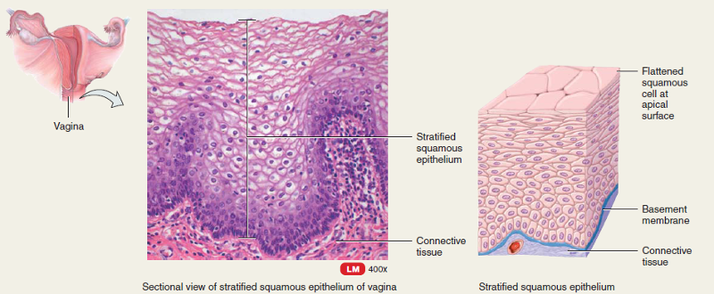
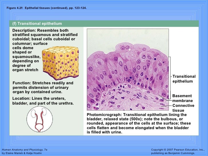
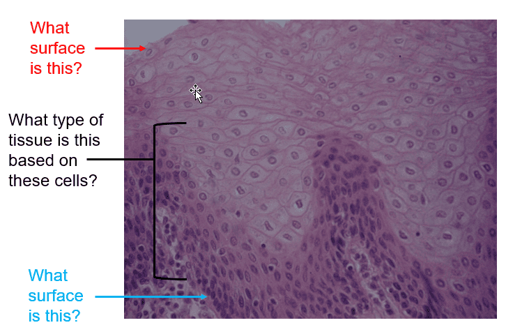
Stratified squamous, epithelium is always flat at the apical end These tissues get harder to tell apart in distended Transnational epithelium, although here, the cells are more uniformly flat, while the flatness is restricted to the apical surface cells in Stratified Squamous Epithelium, unkertinized. The cheek individual squamous cells from the surface of the epithelium can be removed These surface cells are squamous (flat) in shape These cells are part of the nonkeratinized stratified squamous epithelium, but are large and are primarily going to be positioned such that they will be viewed from the “topside” rather than a side view. Simple Squamous Epithelium Definition Simple squamous epithelia are tissues formed from one layer of squamous cells that line surfaces Squamous cells are large, thin, and flat and contain a rounded nucleus Like other epithelial cells, they have polarity and contain a distinct apical surface with specialized membrane proteins.
Simple squamous epithelium ;. Simple Epithelium 1 Simple squamous epithelium is composed of one layer of squamous cells that rests on a basement membrane A surface view shows that the cells have a polygonal outline A side view shows that cells are extremely flattened with flattened nuclei Sites It is characteristically thin to allow material transport across it. Simple squamous epithelium is composed of a single layer of cells that are wider than they are tall This epithelium presents little barrier to passive diffusion and, therefore, lines surfaces across which metabolites or gases can move rapidly This epithelium also lines surfaces that require minimal protection, as shown here Kidney 1000x.
Author Windows User Created Date 09/13/11 Title Simple Squamous Epithelium (top view) Last modified by Windows User. Chief examples are (1) endothelium which covers the internal surface of heart, blood vessels and lymph vessels, (2) mesothelium which lines the pericardial, pleural and peritoneal cavities, and (3) the lining epithelium of the alveoli of the lung. Stratified squamous, epithelium is always flat at the apical end These tissues get harder to tell apart in distended Transnational epithelium, although here, the cells are more uniformly flat, while the flatness is restricted to the apical surface cells in Stratified Squamous Epithelium, unkertinized.
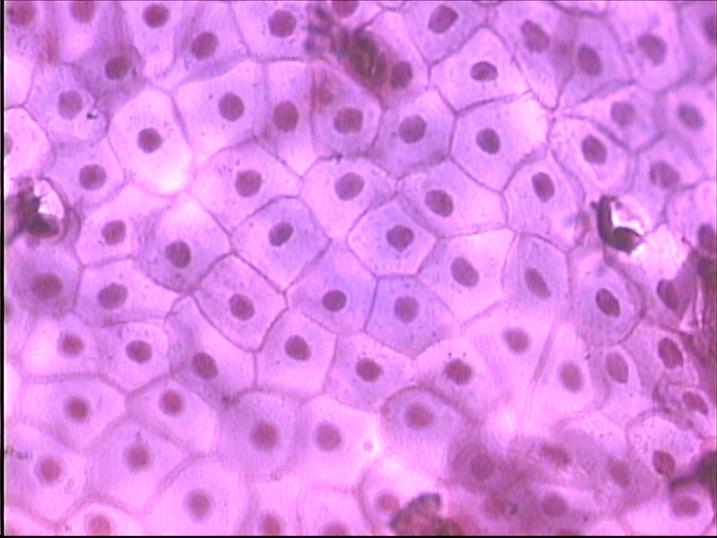
MESOTHELIUM (SIMPLE SQUAMOUS EPITHELIUM) view from surface Stained with silver nitrate 1 nucleus of cell 2 cell borders SIMPLE SQUAMOUS EPITHELIUM Stained with haematoxylin and eosin Nuclei of the epithelial cells are shown with an arrow SIMPLE CUBOIDAL EPITHELIUM. Simple Squamous Epithelium 100X Simple squamous epithelium is constructed of a single layer of flat cells to enable diffusion and filtration of biomolecules (eg, gas exchange within the lung) However, the thin structure results in decreased protection Each cell has a discshaped nuclei Simple Squamous Epithelium 1000X. MESOTHELIUMSIMPLE SQUAMOUS EPITHELIUM, Surface View, 400X, also called "pavement epithelium" These are very flattened cells showing a nucleus, cytoplasm and cell membrane This epithelium forms a thin and often permeable membrane Mesothelium is a special type of simple squamous epithelium that is found in serous membranes lining the pleural.
Simple squamous epithelium mesothelium, surface view, 250x shows simple squamous cells connected (sometimes called pavement epithelium), nuclei, cytoplasm, and cell membranes found in the mesentery simple squamous epithelium stock pictures, royaltyfree photos & images. It consists of a single layer of thin flat, scalelike cells On surface view , the cells have an irregular shape with a slightly serrated border Each cell has a centrally located spherical or oval nucleus In a side view, the cells are so flat that they can only recognize by their elongated nuclei that bulge into. Stratified Squamous Epithelium, Esophagus (40x) Submitted by ghatzipetrou on Fri, 02/16/18 2330 Key L=Lumen.
Where is Mesothelium found?. Renal corpuscle What structure is in the middle of the slide?. Simple Squamous Epithelium 100X Simple squamous epithelium is constructed of a single layer of flat cells to enable diffusion and filtration of biomolecules (eg, gas exchange within the lung) However, the thin structure results in decreased protection Each cell has a discshaped nuclei Simple Squamous Epithelium 1000X.
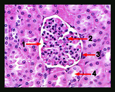
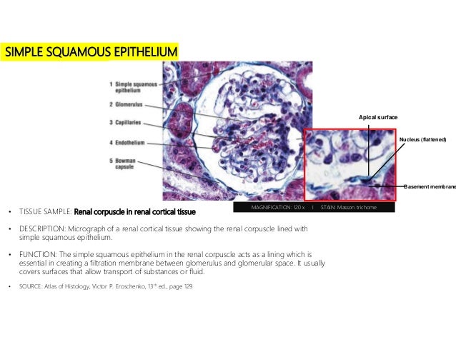
There is a simple squamous epithelial cell nucleus just to the right of the asterisk Simple squamous epithelium, cs (400X) thin section Kidney cortex The arrows in the image point to the nuclei of simple squamous epithelial cells This image was made from a thin section of the kidney at the same magnification as the previous image (400X). This is a surface view of the peritoneum that lines the body cavity of a vertebrate The cementing substance between individual cells is stained black with silver Like a pan of fried eggs, mutual pressure of the cells of this simple squamous epithelium causes them to assume a roughly hexagonal shape. This is a surface view of the peritoneum that lines the body cavity of a vertebrate The cementing substance between individual cells is stained black with silver Like a pan of fried eggs, mutual pressure of the cells of this simple squamous epithelium causes them to assume a roughly hexagonal shape.
They lie on a very thin basement membrane The side of an epithelial layer opposite the basement membrane is called its free surface See simple squamous and simple cuboidal epithelia on the KIDNEY slide in Chapter 10;. Under microscope, magnification 600X View in microscopic of ductal cell carcinoma, adenonocarcinoma from human breast cancer, tissue section by H and E stainPathology diagnosisMedical concept Under microscope, magnification 600X simple squamous epithelium stock pictures, royaltyfree photos & images. Simple squamous epithelium (mesothelium) surface view, 250x shows shows squamous cells connected (sometimes called pavement epithelium), nuclei, cytoplasm, cell membrane this epithelium is found in the mesentery squamous epithelial cells stock pictures, royaltyfree photos & images.
The cheek individual squamous cells from the surface of the epithelium can be removed These surface cells are squamous (flat) in shape These cells are part of the nonkeratinized stratified squamous epithelium, but are large and are primarily going to be positioned such that they will be viewed from the “topside” rather than a side view. Simple squamous epithelium consists of a single layer of thin, flat, scalelike cells On surface view (Figs 23A and 24), the cells have an irregular shape with a slightly serrated border They fit together to form a continuous sheet A spherical to oval nucleus is located near the center of the cell. The cheek individual squamous cells from the surface of the epithelium can be removed These surface cells are squamous (flat) in shape These cells are part of the nonkeratinized stratified squamous epithelium, but are large and are primarily going to be positioned such that they will be viewed from the “topside” rather than a side view.
A stratified epithelium is more than one layer of cells thick A pseudostratified epithelium is really a specialized form of a simple epithelium in which there appears at first glance to be more than one layer of epithelial cells, but a closer inspection reveals that each cell in the layer actually extends to the basolateral surface of the. “In the surface view of simple squamous epithelium, cell looks polygonal shaped with serrated border Nucleus is oval or spherical shaped and present in the center of the cell” Location of simple squamous epithelium You will find simple squamous epithelium on –. Simple squamous epithelium is of common occurrence in the body;.
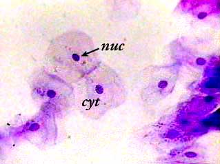
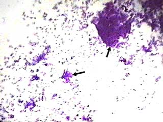
Simple squamous epithelium, isolated (400x) Buccal mucosal In the center of this image are two simple squamous epithelial cells that are still attached to each other Notice that the location of the nucleus (nuc) is in the center of the cell It is surrounded by the much paler cytoplasm (cyt). It consists of a single layer of thin flat, scalelike cells On surface view , the cells have an irregular shape with a slightly serrated border Each cell has a centrally located spherical or oval nucleus In a side view, the cells are so flat that they can only recognize by their elongated nuclei that bulge into. Squamous epithelial cells are typically discrete in cross section, appearing as thin lines with a protrusion in the nucleus A simple squamous epithelium is so thin that it is barely visible by light microscopy A stratified squamous epithelium is quite thick, with squamous cells on the surface coating deeper layers of higher cells.
Stratified squamous epithelium is the most common type of stratified epithelium in the human body The apical cells appear squamous, whereas the basal layer contains either columnar or cuboidal cells The top layer may be covered with dead cells containing keratin The skin is an example of a keratinized, stratified squamous epithelium. Simple Epithelium 1 Simple squamous epithelium is composed of one layer of squamous cells that rests on a basement membrane A surface view shows that the cells have a polygonal outline A side view shows that cells are extremely flattened with flattened nuclei Sites It is characteristically thin to allow material transport across it. Simple squamous epithelium is of common occurrence in the body;.
Simple Epithelium 1 Simple squamous epithelium is composed of one layer of squamous cells that rests on a basement membrane A surface view shows that the cells have a polygonal outline A side view shows that cells are extremely flattened with flattened nuclei Sites It is characteristically thin to allow material transport across it. SIMPLE SQUAMOUS EPITHELIUM (Mesothelium) Surface View, 250X Shows shows squamous cells connected (sometimes called pavement epithelium), nuclei, cytoplasm, cell membrane This epithelium is found in the mesentery stock photo. The simple squamous epithelium forming the mesothelium facilitated the movement of the viscera and the active transport of fluid by the process of pinocytosis 3 Secretion Some cells in the simple squamous epithelium are also known to produce some fluids like the mucus that acts as a lubricating agent against the inevitable friction.
This is a diagram of a surface view of a simple squamous epithelium You will not see such a preparation This is for illustration only A piece of epithelium, spread on a slide is treated with a silver salt which brings out the cell membrane and cell boundaries Since no other stain is used, there is no 'colour' in the cytoplasm or in the nucleus. Mesothelium (Surface view) Simple squamous epithelium Name the epithelium?. The simple squamous epithelium forming the mesothelium facilitated the movement of the viscera and the active transport of fluid by the process of pinocytosis 3 Secretion Some cells in the simple squamous epithelium are also known to produce some fluids like the mucus that acts as a lubricating agent against the inevitable friction.
Stratified Squamous Epithelium, Esophagus (40x) Submitted by ghatzipetrou on Fri, 02/16/18 2330 Key L=Lumen. Projections of a columnar cell that increase its surface area or assist in moving substances along the cell surface microvilli/cilia Pseudostratified columnar epithelium Simple squamous Stratified Squamous Epithelium A Cilia B Goblet cell. Simple squamous epithelium mesothelium, surface view, 250x shows simple squamous cells connected (sometimes called pavement epithelium), nuclei, cytoplasm, and cell membranes found in the mesentery simple squamous epithelium stock pictures, royaltyfree photos & images.
Simple Squamous Epithelium, 40X Submitted by ipolunina on Wed, 02/21/18 1338 Read more about Simple Squamous Epithelium, 40X;.
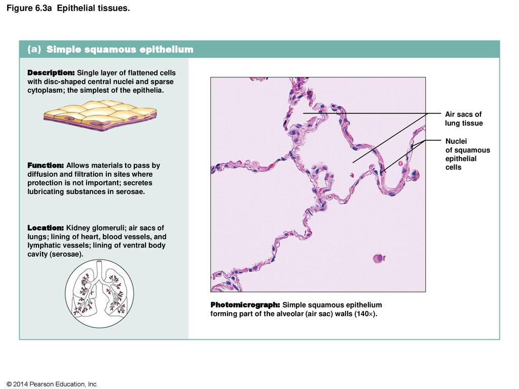
Figure 6 1 Classification Of Epithelia Ppt Download
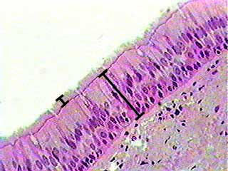
Pseudostratified Ciliated Epithelium
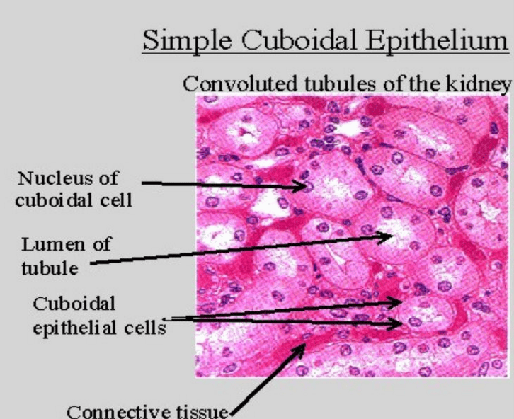
Print A P Tissues Ch 4 Flashcards Easy Notecards
Simple Squamous Epithelium Surface View 1000x のギャラリー
I E All
Www Studocu Com En Us Document Eastern Kentucky University Human Physiology Lecture Notes Ch 4 Histology Lecture Over Tissue Types View

95 Simple Columnar Epithelium Photos And Premium High Res Pictures Getty Images
Stratified Squamous Keratinized Epithelium

Epithelial David Fankhauser

30 Epithelium Ideas Histology Slides Anatomy And Physiology Human Anatomy And Physiology
Histology Flashcards Chegg Com

Epithelial David Fankhauser
Q Tbn And9gctzu7fd7f 63uvsuhii Iwznc54mlzqmr8fup1orkbsues3tngw Usqp Cau
Q Tbn And9gcsz7vu4vzv64expinjqs4vhk M3odj3fjmzmtlku3kgqwxp3azs Usqp Cau

Histology Lab With Answers

Lab 2 Epithelial Tissue Histology

Histology Lab With Answers

Chapter 4 Tissue Level Of Organization Ppt Video Online Download

Stratified Cuboidal Epithelium Composed Of Several Layers Of Cuboidal Download Scientific Diagram
Simple Squamous Epithelium Isolated

Simple Squamous Epithelium
Print A P Tissues Ch 4 Flashcards Easy Notecards

Histology Series

Simple Squamous Epithelium Kit Ng Ph D

Figure 6 1 Classification Of Epithelia Ppt Download
Tissues
Simple Squamous 1
Terminology Tissues Lab Festival Flashcards Easy Notecards

Learning Objectives Keywords Pre Lab Reading Pre Lab Quiz Slides Virtual Microscope Pathology Quiz Epithelia Lab Learning Objectives Explain The Ways In Which Epithelia Are Classified Distinguish Between Simple Stratified And Pseudostratified

Histolab2 Htm

Histology Lab With Answers
Q Tbn And9gct7egvdok63lxw Suvrh1qvsnsvgadjy3bxyyazzgreqvz96p Usqp Cau

Chapter 4 Tissue Level Of Organization Ppt Video Online Download
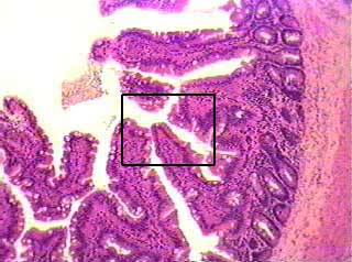
Simple Columnar Epithelium

Lab Exercises 2 3 Flashcards Quizlet

Chapter 1 Page 7 Histologyolm
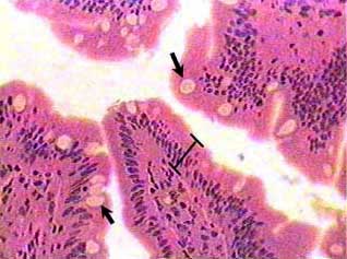
Simple Columnar Epithelium

Learning Objectives Keywords Pre Lab Reading Pre Lab Quiz Slides Virtual Microscope Pathology Quiz Epithelia Lab Learning Objectives Explain The Ways In Which Epithelia Are Classified Distinguish Between Simple Stratified And Pseudostratified

95 Simple Columnar Epithelium Photos And Premium High Res Pictures Getty Images

Histolab2 Htm
I E All

Simple Squmous Epithelium C S

Microscopic Histology Images Epithelial Tissue

New Page 2

Ap1 Histology Slides Flashcards Quizlet
Http Images Pcmac Org Sisfiles Schools Pa Greenvillearea Greenvillejrsrhigh Uploads Presentations Chapt04 Apr Lecture 7bsisecf8f8 7d Pdf

Ctr Histology Study Flashcards Quizlet
I E All
Stratified Squamous Keratinized Epithelium

Epithelial David Fankhauser

Stratified Squamous Epithelium

Histolab2 Htm

Histolab2 Htm

What Type Of Epithelium Lines The Inner Surface Of This Type Of Epithelium In This Organ

Anatomy Types Of Epithelium You Ll Remember Quizlet
Surface Of Simple Squamous Epithelium Silver Staining Histology Slides Buy Microscope Slides Histology Slides Histology Prepared Slides Product On Alibaba Com
Massasoit Instructure Com Files Download Download Frd 1 Verifier J66jcmre6tluf6g4x5n3vslnfwaqlore1uw3c2ku

Types Of Epithelial Tissue Flashcards Quizlet

Microscopic Histology Images Epithelial Tissue

Histology Epithelial Tissue Flashcards Quizlet
Epithelia The Histology Guide

Epithelium Web Lab

Lab Manual Exercise 1
Epithelium Web Lab

Lab Manual Exercise 1

Solved Figure 6 5 Lumen Of Kidney Tubules Glomerulus Simp Chegg Com
Q Tbn And9gcskx04lcbk Fptlwmgrku8pxxpte1lwkn873papzge Ysfruacx Usqp Cau

Volner Ilab3 Labex6 Docx Lab Activity 1 Microscopic Examination Of Epithelia 1 Examine A Prepared Microscope Slide Of A Whole Mount Of Mesothelium Use Course Hero
Simple Squamous Epithelium Isolated

Histology Slides Flashcards Quizlet

Learning Objectives Keywords Pre Lab Reading Pre Lab Quiz Slides Virtual Microscope Pathology Quiz Epithelia Lab Learning Objectives Explain The Ways In Which Epithelia Are Classified Distinguish Between Simple Stratified And Pseudostratified
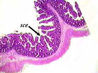
Simple Columnar Epithelium
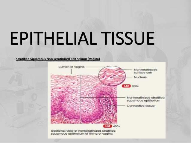
I E All
Http Learning Hccs Edu Faculty M Bracamonte Biol 2401 Ap 1 course material Ap 1 Lab Handouts Tissues Tissues Micrographs At Download File

Simple Squamous Epithelium Structure Functions Examples

Learning Objectives Keywords Pre Lab Reading Pre Lab Quiz Slides Virtual Microscope Pathology Quiz Epithelia Lab Learning Objectives Explain The Ways In Which Epithelia Are Classified Distinguish Between Simple Stratified And Pseudostratified
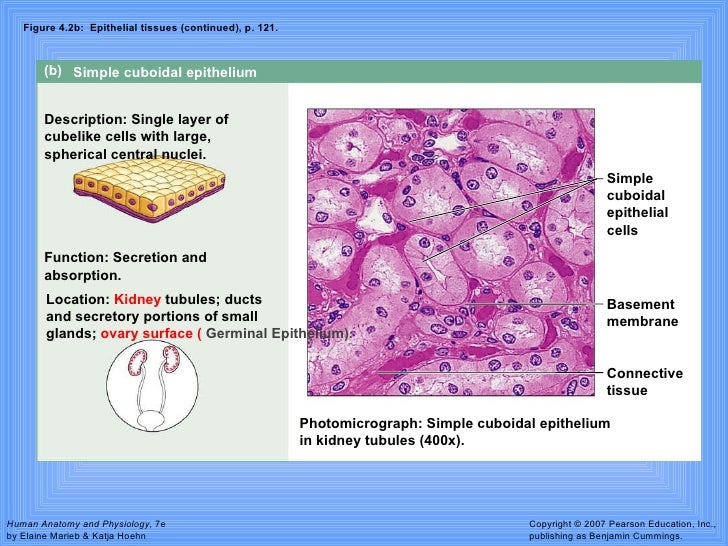
Tissues
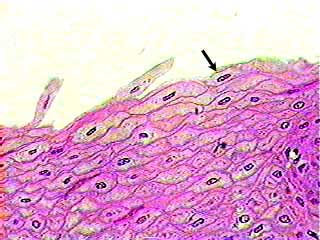
Stratified Squamous Non Keratinized Epithelium

Lab 2 Epithelial Tissue Histology

Lab Practical Tissue Slides Base Flashcards Quizlet

Simple Squamous Epithelium 40x Annotated Histology

Cheek Cells Under The Microscope Youtube Animal Cell Microscopic Things Under A Microscope

Volner Ilab3 Labex6 Docx Lab Activity 1 Microscopic Examination Of Epithelia 1 Examine A Prepared Microscope Slide Of A Whole Mount Of Mesothelium Use Course Hero
Http Images Pcmac Org Sisfiles Schools Pa Greenvillearea Greenvillejrsrhigh Uploads Presentations Chapt04 Apr Lecture 7bsisecf8f8 7d Pdf
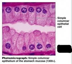
Terminology Tissues Lab Festival Flashcards Easy Notecards

Stratified Squamous Epithelium With Several Layers Of Squamous Cells Download Scientific Diagram

Figure 6 1 Classification Of Epithelia Ppt Download
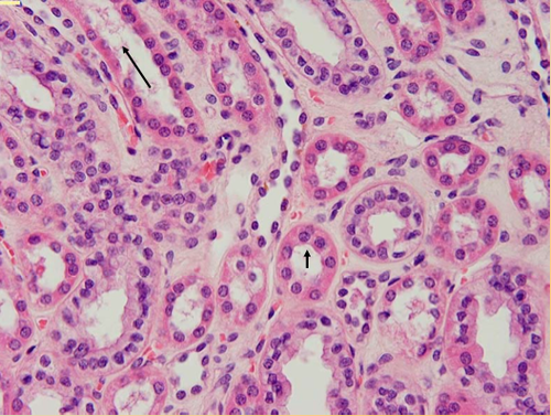
Histology Slides Flashcards Quizlet

Epithelial David Fankhauser

Ap1 Histology Slides Flashcards Quizlet

Simple Squmous Epithelium C S

Simple Squamous Epithelium Isolated

Epithelial Tissue By Kourtney Cooper

Histology Lab With Answers
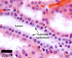
Epithelia The Histology Guide

Solved Figure 6 5 Lumen Of Kidney Tubules Glomerulus Simp Chegg Com

Surface Papillary Projections Lined By A Parakeratotic Acanthotic Download Scientific Diagram

Histolab2 Htm

Stratified Squamous Epithelium

Simple Squamous Epithelium Surface View Endothelium Of Vein A And Download Scientific Diagram
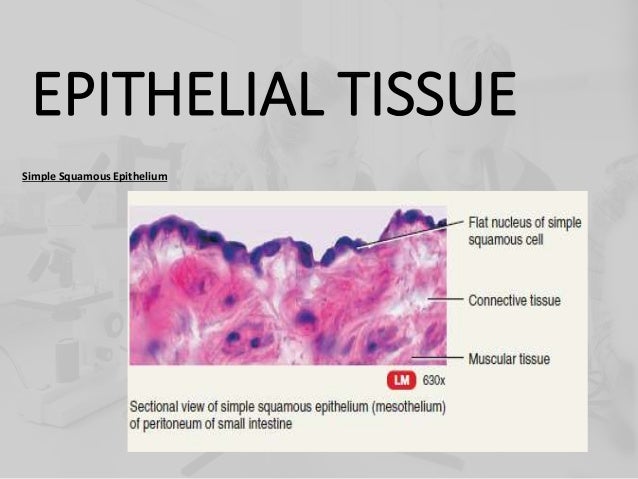
I E All

A P Lab Exam 1 Tissues Pictures Flashcards Quizlet
Http Histologyguide Org About Us Sorenson Atlas Of Human Histology Chapters 1 And 14 Pdf
A P 1 Unit 6 Epithelium Nervous And Muscle Tissues Flashcards Easy Notecards
Epithelial Tissues Ppt Download